
Mobile Source
Researching student: *hiring*
Most recent publications: Time-resolved imaging of laser-induced phase transitions in free silver nanoclusters
Introduction
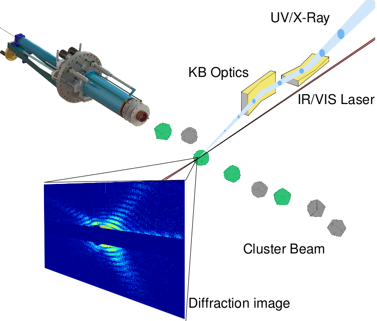
Control of nanoscale matter proved promising in a vast range of applications, be it medical purposes such as cancer therapy, chemical analysis and catalysis, or nonlinear optical processes (see e.g. [1]). Recently it has been shown feasible to image the 3D structure of single metal nanoparticles in a diffraction-before-destruction setup at the free electron laser FLASH in Hamburg [2]. Extending this technique into a pump-probe probe experiment will yield insight into the behavior of nanoparticles at sub-nanosecond timescale. Using an IR laser to start interesting behavior and a probe X-Ray pulse to obtain a diffraction image of the state of the cluster at a given time. In this way we will obtain "flicker-book movies" of melting clusters. Such yield insights into the electron dynamics of the cluster at extreme conditions.
Challenges
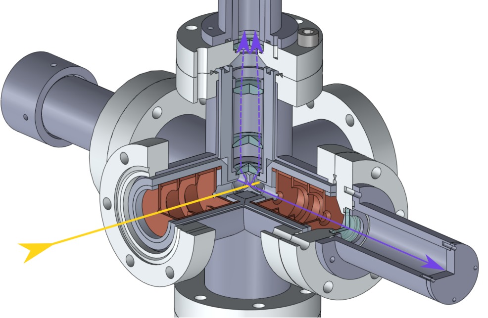
Due to the low scattering cross sections of nanoclusters, high photon fluxes are necessary. At FLASH, a beam waist as narrow as 10 microns is possible. With a cluster beam of one millimeter in diameter, the interaction volume resembles a cylinder of 10−13 cubic meters. To have a single particle in this volume about every 10 shots (X-Ray pulses are short enough (fs) that movement of the clusters can be neglected), the density needs to be in the order of 1012 clusters per cubic meter or 106 cm-3. Assuming single-charged clusters, this amounts to a cluster current of 0.3 A (given a realistic cluster velocity of 150m/s and 4mm diameter of the clusterbeam). A further constraint for such experiments pertains to the size of the nanoclusters. Because the scattering cross section decreases with higher photon energy, larger clusters are preferable. Their special features are larger and thus better resolved with lower energy photons. At FLASH, photon energies of up to 310eV are achievable. Due to this limitation, structures smaller than a few nanometers can not be resolved. We aim to produce clusters larger than 30nm, preferably 90nm as here feasibility has been shown [2].
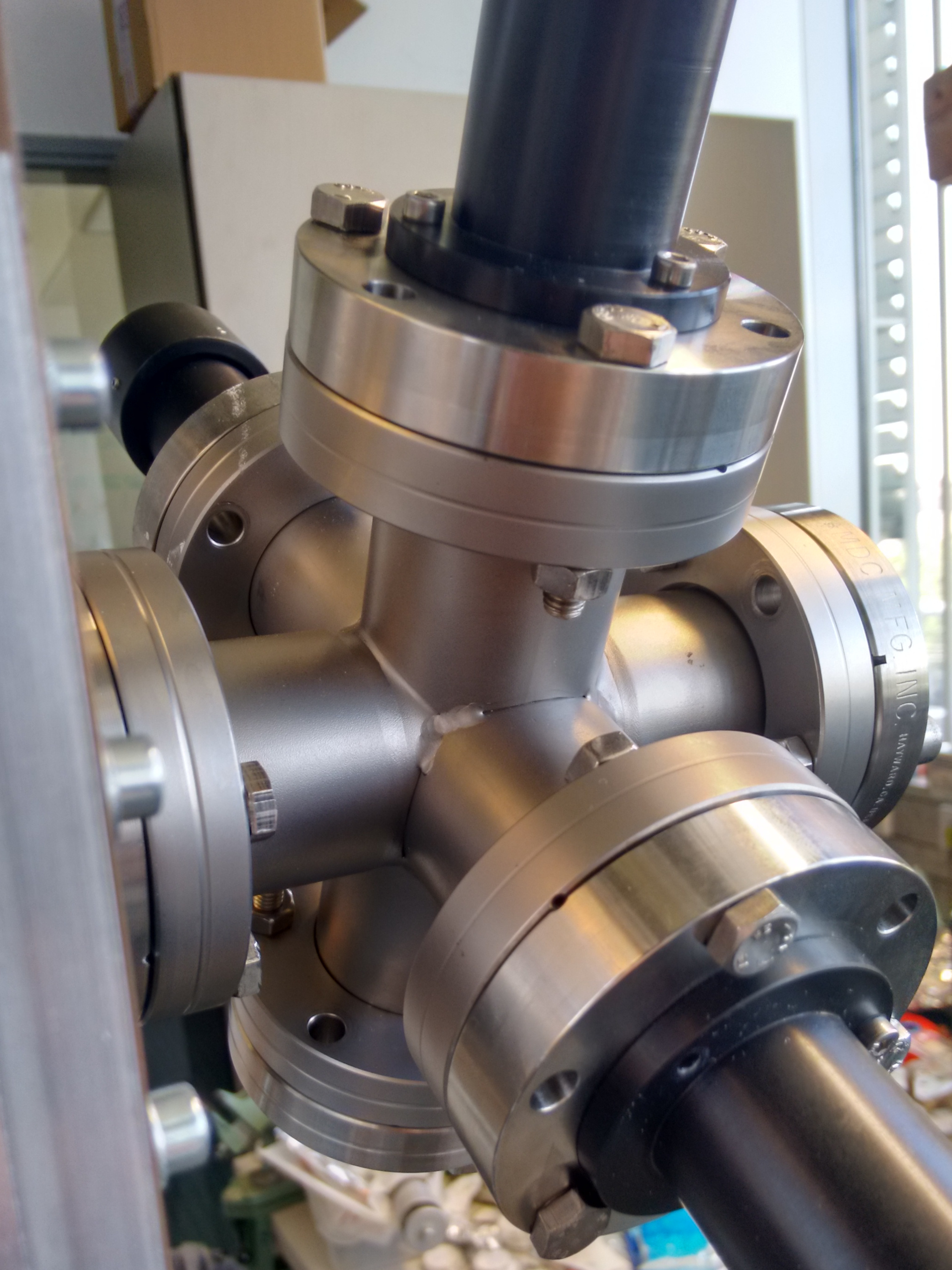
Current Work
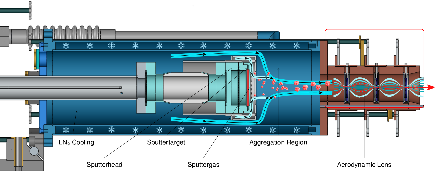
Precise control of size in the synthesis of various nanoparticles is well established. Large clusters from gas aggregation sources have been observed, however, their formation mechanism is not well understood. Great progress towards the production of large clusters has been made, however generation of clusters larger that 20nm in diameter remains difficult.
Microscopy
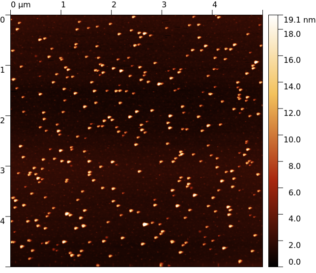
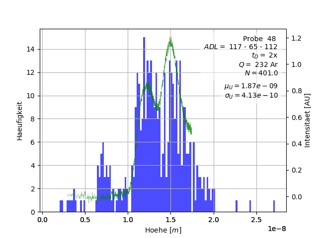
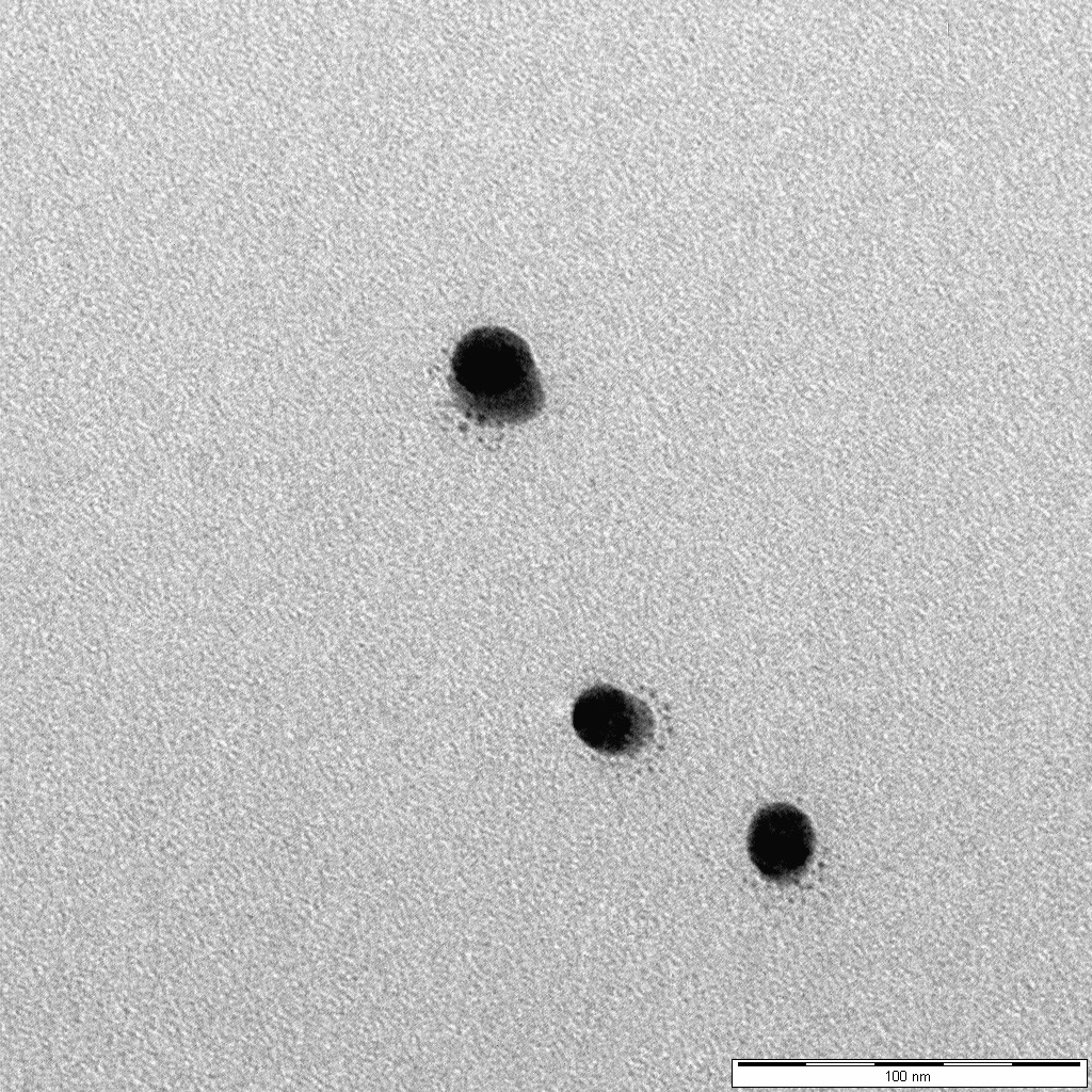
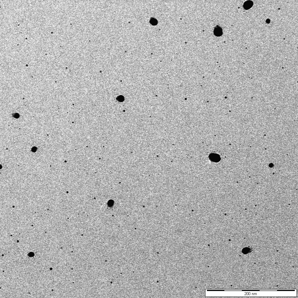
To verify the results above, clusters have been deposited on silicon substrates and TEM grids.

Mie scattering
To optimize source parameters for large clusters in real-time, a detector exploiting Mie scattering has been devised. As clusters approach sizes larger than 20nm, scattering of in-plane polarized 405nm laser-light at 90° with respect to the incoming laser light drastically increases. This, in principle, allows us to detect even small amounts of large clusters in the cluster beam in real-time.
Forthcoming Research
- Real-time detection of large clusters exploiting Mie scattering to deepen our understanding of the conditions leading to large clusters.
- Time-resolved pump-probe on metal clusters.
- Vincenzo Amendola, Roberto Pilot, Marco Frasconi, Onofrio M Maragò, Maria Antonia Iatì Surface plasmon resonance in gold nanoparticles: a review J Phys Condens Matter. 2017 May 24;29(20):203002
- Ingo Barke, Hannes Hartmann, Daniela Rupp, Leonie Flückiger, Mario Sauppe, Marcus Adolph, Sebastian Schorb, Christoph Bostedt, Rolf Treusch, Christian Peltz, Stephan Bartling, Thomas Fennel, Karl-Heinz Meiwes-Broer & Thomas Möller The 3D-architecture of individual free silver nanoparticles captured by X-ray scattering Nature Communications volume 6, Article number: 6187 (2015)
- Samuel Kellerer. Charakterisierung einer Magnetron-Clusterquelle durch Clusterdeposition, 2017.